Can I have an x-ray taken with brackets?


Wearing metallic braces may generate the doubt as to whether it is possible or even dangerous to have an x-ray test done. In this article we shed light on this matter and go into depth regarding x-rays in the field of odontology.
How do x-rays work?
Radiology is a medical specialism for taking images of the inside of the body using different physical agents such as x-rays or magnetic fields. These images allow, together with the remaining signs and symptoms and the professional’s assessment, a diagnosis of the pathology to be made, and a possible prognosis and treatment.
X-rays are used to obtain images both in the medical and dental field. These images are obtained when an emission of light crosses the body and hits a sensor or an x-ray film on the opposite side. The shadow that is protected on the film enables the professional to see the structures covered by the skin.
The professionals may distinguish the normal tissue from the anomalies, deciphering the difference between the clear and dark patterns that appear in the x-ray.
Are x-rays necessary for an orthodontic treatment?
In orthodontics, panoramic x-rays, just like the lateral skull teleradiography, are fundamental to diagnose problems. Without them, the orthodontist cannot diagnose adequately nor prepare a correct treatment plan. Aside from seeking possible pathologies, orthodontists also observe the position and form of the teeth and jaw.
Assessing the roots of the teeth is also very important when preparing a correct orthodontic treatment plan. Their positioning and length can determine the amount of time the patient can be using brackets.
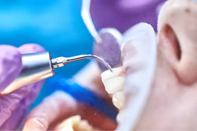
But can x-rays be taken when wearing brackets?
Once the treatment has begun, with the brackets now cemented to the teeth, it is necessary to carry out x-rays both to monitor the progress of the treatment and to assess the appearance of new pathologies as could be the presence of tooth decay.
Conventional x-rays
Therefore, if during the orthodontic brackets treatment, it is necessary to carry out x-rays or tomographies, both to assess possible pathologies in the mouth cavity and in other adjacent areas of the head or neck, there is no problem using x-rays.
Resonances and tomographies
In the case of magnetic resonances, in which ionising radiation is not used as in computerised tomographies or in conventional x-rays, rather radiowaves and magnetic fields are used to generate anatomical images of a part of the body, the patient must be free from extractable metallic objects such as coins and keys, since metallic objects can cause safety risks. Some metallic objects cannot be easily removed, such as orthopaedic devices or implantable medical devices, including metallic brackets, retainers and certain palate expanders.
In principle, with this type of device, the patient can have magnetic resonance tests, since the brackets are sufficiently attached to the teeth and it has been demonstrated that they are safe.
Even so the radiologist may ask a patient to request that their odontologist removes the specific device to allow the patient to have a magnetic resonance in a safe manner without worrying that the patient will suffer an injury
Possible effects of brackets on x-rays
Firstly although they can be safe, these devices may cause artefacts in the images generated. These artefacts predominantly occur in the facial and orbital regions, although the artefacts can also be extended including areas of the brain and cervical spine.
Hence on occasions the radiologist requests the orthodontist to withdraw the brackets from the patient to allow both him and the patient’s doctor to correctly assess structures close to the face that can seem darkened by the orthodontic items, and thereby not interfere in the diagnosis.